Microphysiological systems and organ-on-a-chip models
Tumours are shaped by a dynamic tissue environment in which blood and lymphatic vessels regulate oxygen and nutrient supply, drug delivery, fluid drainage, and cell trafficking. Reproducing these transport processes is one of the central challenges in building microphysiological systems that remain viable, functional, and biologically relevant over time.
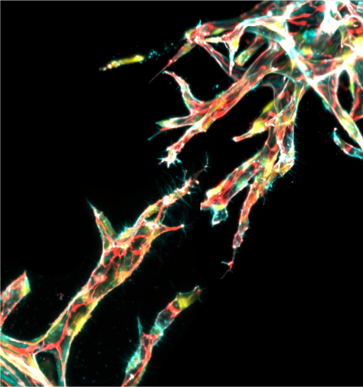
Our focus is on developing microfluidic models that integrate perfused vascular and lymphatic networks with tumour spheroids or organoids, stromal cells, and engineered extracellular matrices. These systems will provide control over flow, vascular permeability, tissue mechanics, and local chemical gradients while enabling the study of how tumours and their surrounding tissues change over time. The resulting platforms will be used to investigate tumour–vascular interactions, angiogenesis, lymphatic function, barrier transport, invasion, metastasis, drug delivery, and therapeutic response. By bringing key elements of the tumour microenvironment into a controlled setting, the goal is to create more informative models of cancer progression and patient-relevant treatment strategies.
.png)
Biomanufacturing
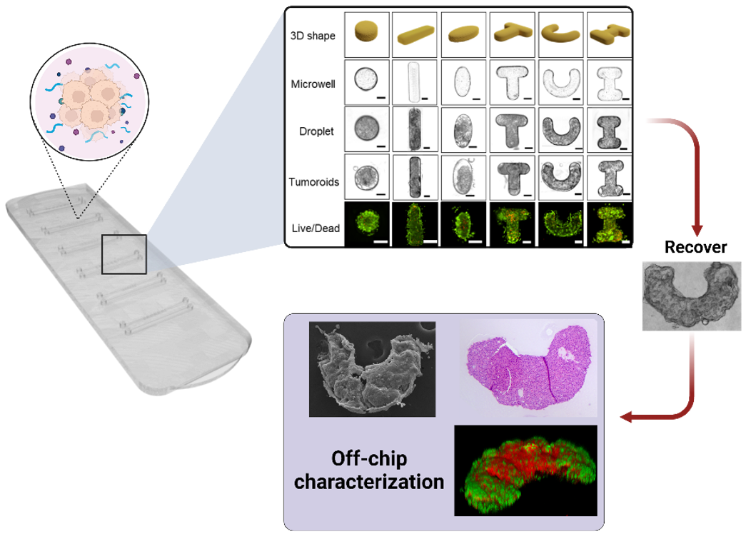
Biomanufacturing approaches provide control over the composition, architecture, and function of living tissue models. Through narrative engineering, we design the spatial, biochemical, and mechanical environments that guide stochastic cellular behaviors toward more organized and physiologically meaningful tissue outcomes. Rather than relying on self-organization alone, these approaches help direct how cells interact, assemble, and evolve over time.
Using droplet-based microfluidics and 3D bioprinting, we generate shape-defined cellular building blocks and arrange them into larger, structured tissue constructs. These methods provide control over tissue geometry, cellular organization, and matrix composition while allowing dynamic cues to be incorporated during formation. By combining modular assembly with mechanical stimulation, we aim to guide angiogenesis and vascular network formation and create reproducible, vascularized models for disease research and therapeutic testing.
Computational approaches
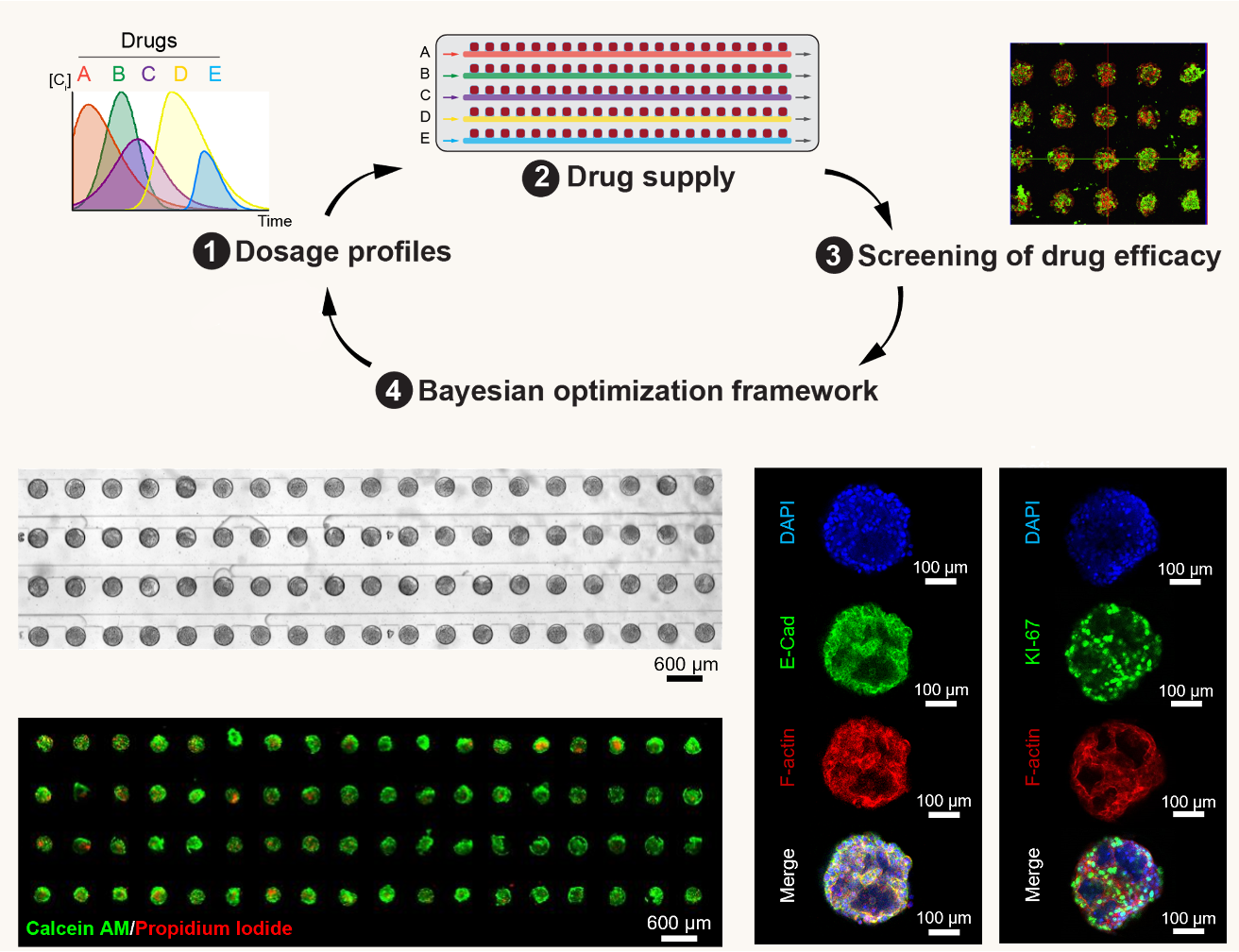
Computational fluid dynamics, digital twins, automation, and machine learning are used to make microphysiological systems more predictive and useful for therapeutic testing. Digital-twin models of microfluidic platforms help describe how device geometry, flow conditions, and transport processes influence the local delivery of drugs, nutrients, and biochemical signals. These models can guide device design before fabrication and help establish controlled, physiologically relevant experimental conditions.
Combined with automated microfluidics, computational approaches can support systematic exploration of complex treatment spaces. Data-driven models help analyze large experimental datasets and identify informative conditions across drug dose, timing, sequence, and combination strategies. The long-term goal is to develop patient-relevant platforms that can support individualized drug screening while reducing the experimental burden required to reach clear decisions.